Home
/ Medial Femoral Condyle Flap : Https Www Plasticsurgery Theclinics Com Article S0094 1298 16 30178 X Pdf, The medial femoral condyle has become an area of increased interest given the versatility it allows in harvesting the bone.
Medial Femoral Condyle Flap : Https Www Plasticsurgery Theclinics Com Article S0094 1298 16 30178 X Pdf, The medial femoral condyle has become an area of increased interest given the versatility it allows in harvesting the bone.
Medial Femoral Condyle Flap : Https Www Plasticsurgery Theclinics Com Article S0094 1298 16 30178 X Pdf, The medial femoral condyle has become an area of increased interest given the versatility it allows in harvesting the bone.. The saphenous branch of the descending genicular artery supplies the medial femoral condyle skin flap. Design, harvest, and inset of trimmed great toe for thumb reconstruction in mutilated hand The medial femoral condyle has become an area of increased interest given the versatility it allows in harvesting the bone. 1,2 the benefit of this type of flap is the viability of the bone which favors primary ossification and increases bone density. We had a flap tear of the articular cartilage based medially and coming out of the notch and the entire area of the femoral condyle.
The medial femoral condyle flap is used for treatment of nonunions with or without intercalary bone loss. The use of the medial femoral condyle free flap (mfcf) is a versatile option for the treatment of upper and lower extremity nonunions and reconstructive procedures associated with bone loss or avascular necrosis. The vascularized medial femoral condyle corticoperiosteal flap provides an additional treatment option for the management of humeral nonunions. Chondral flap (cartilage separates from the bone and moves like a door with a hinge at one end) chondral fracture (cartilage separates from the bone and floats free) chondral lesions may be degenerative (a wear and tear problem) or traumatic (caused by an injury such as falling on the knee, jumping down, or rapidly changing direction while. The medial femoral condyle (mfc) flap, which incorporates cortical bone, cancellous bone, periosteum, muscle, tendon, cartilage, and skin, is well suited for complex reconstructions2.
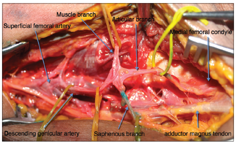
1 from Vascularized medial femoral condyle corticoperiosteal flaps for the treatment of recalcitrant humeral nonunions. The aim of this report is to present the functional and aesthetical outcomes of a reconstruction of the nose after rhinectomy, using the medial femoral condyle free flap associated with the forehead flap. Figure 55.2 (a) schematic of the anatomy of the descending genicular artery (dga) and superomedial genicular arterial system. The medial femoral condyle (mfc) flap, which incorporates cortical bone, cancellous bone, periosteum, muscle, tendon, cartilage, and skin, is well suited for complex reconstructions2. Most reported uses have been without a skin segment, but this flap can provide a skin component supplied by the saphenous artery branch (sab) of the descending genicular artery (dga) pedicle. The use of the medial femoral condyle free flap (mfcf) is a versatile option for the treatment of upper and lower extremity nonunions and reconstructive procedures associated with bone loss or avascular necrosis. A retrospective review was completed of all mfc flaps used in the foot and ankle over the past 5 years. Design, harvest, and inset of trimmed great toe for thumb reconstruction in mutilated hand
A composite osteomusculocutaneous free flap from the medial femoral condyle for reconstruction of complex defects.
The medial femoral condyle (mfc) flap has emerged as a popular source of vascularized corticocancelous bone. There was no medial meniscus tear. Vascularized medial femoral condyle corticoperiosteal flaps for the treatment of recalcitrant humeral nonunions. The use of the medial femoral condyle free flap (mfcf) is a versatile option for the treatment of upper and lower extremity nonunions and reconstructive procedures associated with bone loss or avascular necrosis. The vascularized medial femoral condyle corticoperiosteal flap provides an additional treatment option for the management of humeral nonunions. Kakar s., duymaz a., steinmann s., et. Pain is often located along the inner (medial) aspect of the knee. This flap can provide a versatile source of vascularized bone with limited donor site morbidity and requires minimal time for flap harvest. The medial femoral condyle vascularized bone flap is a versatile option, with simple dissection and relatively constant anatomy for reconstruction of musculoskeletal injuries.its irrigation from the descending genicular artery (or the superior medial genicular artery if the descending genicular artery is absent) allows transfer of the. We had a flap tear of the articular cartilage based medially and coming out of the notch and the entire area of the femoral condyle. The aim of this report is to present the functional and aesthetical outcomes of a reconstruction of the nose after rhinectomy, using the medial femoral condyle free flap associated with the forehead flap. The medial femoral condyle (mfc) flap, which incorporates cortical bone, cancellous bone, periosteum, muscle, tendon, cartilage, and skin, is well suited for complex reconstructions2. We present a series of cases demonstrating the versatility of the mfc flap in complex foot and ankle pathology.
A composite osteomusculocutaneous free flap from the medial femoral condyle for reconstruction of complex defects. We had a flap tear of the articular cartilage based medially and coming out of the notch and the entire area of the femoral condyle. Vascularized medial femoral condyle corticoperiosteal flaps for the treatment of recalcitrant humeral nonunions. The flaps of the medial femoral condyle (mfc) are based on the geniculate arterial system, which is a branch of the femoral artery and is located medially in the thigh and knee. For these specific circumstances we have opted to use the the medial femoral condyle corticoperiosteal (mfc) flap.
Vascularised Corticoperiosteal Grafts From The Medial Femoral Condyle For Difficult Non Unions Of The Upper Limb Sciencedirect from ars.els-cdn.com The benefit of this type of flap is the viability of the bone which favors primary ossification and increases bone density. 1,2 the benefit of this type of flap is the viability of the bone which favors primary ossification and increases bone density. Design, harvest, and inset of trimmed great toe for thumb reconstruction in mutilated hand The medial femoral condyle has become an area of increased interest given the versatility it allows in harvesting the bone. There was no medial meniscus tear. A composite osteomusculocutaneous free flap from the medial femoral condyle for reconstruction of complex defects. I hope that the video is descrip. The aim of this report was to develop and evaluate a new method for full thickness total/subtotal nose reconstruction using the medial femoral condyle free flap (mfcff) in combination with a paramedian forehead flap.
We present a series of cases demonstrating the versatility of the mfc flap in complex foot and ankle pathology.
Most reported uses have been without a skin segment, but this flap can provide a skin component supplied by the saphenous artery branch (sab) of the descending genicular artery (dga) pedicle. The aim of this report was to develop and evaluate a new method for full thickness total/subtotal nose reconstruction using the medial femoral condyle free flap (mfcff) in combination with a paramedian forehead flap. The medial femoral condyle flap is used for treatment of nonunions with or without intercalary bone loss. 1 (see video, supplemental digital content 1, which displays a dissection of mfc flap. Lateral femoral condyle flap chetan s. Design, harvest, and inset of trimmed great toe for thumb reconstruction in mutilated hand We put a shaver and a punch in and trimmed down the medial meniscus. A retrospective review was completed of all mfc flaps used in the foot and ankle over the past 5 years. 1,2 the benefit of this type of flap is the viability of the bone which favors primary ossification and increases bone density. Figure 55.2 (a) schematic of the anatomy of the descending genicular artery (dga) and superomedial genicular arterial system. The medial femoral condyle vascularized bone flap is a versatile option, with simple dissection and relatively constant anatomy for reconstruction of musculoskeletal injuries.its irrigation from the descending genicular artery (or the superior medial genicular artery if the descending genicular artery is absent) allows transfer of the. A composite osteomusculocutaneous free flap from the medial femoral condyle for reconstruction of complex defects. The medial femoral condyle (mfc) free flap has recently been introduced, which consists of corticoperiosteal bone.
A composite osteomusculocutaneous free flap from the medial femoral condyle for reconstruction of complex defects. He subsequently underwent revision surgery with resection of the pseudarthrosis, plate fixation, and establishment of a vascularized medial femoral condyle (mfc) flap to ensure bone union. The medial femoral condyle vascularized bone flap is a versatile option, with simple dissection and relatively constant anatomy for reconstruction of musculoskeletal injuries.its irrigation from the descending genicular artery (or the superior medial genicular artery if the descending genicular artery is absent) allows transfer of the. Three months after the revision surgery, a radiographic bone union was achieved, and no symptoms were observed for one year after the operation. Kakar s., duymaz a., steinmann s., et.
Anatomic Variability Of The Vascularized Composite Osteomyocutaneous flap From The Medial Femoral Condyle An Anatomical Study from oaepublishstorage.blob.core.windows.net The benefit of this type of flap is the viability of the bone which favors primary ossification and increases bone density. The use of the medial femoral condyle free flap (mfcf) is a versatile option for the treatment of upper and lower extremity nonunions and reconstructive procedures associated with bone loss or avascular necrosis. The medial femoral condyle has become an area of increased interest given the versatility it allows in harvesting the bone. 1 (see video, supplemental digital content 1, which displays a dissection of mfc flap. Medial femoral condyle flap with skin paddle matthew l. The medial femoral condyle (mfc) free flap has recently been introduced, which consists of corticoperiosteal bone. Pain is often located along the inner (medial) aspect of the knee. Medial femoral condyle free flap for head and neck reconstruction the mfc flap has been successfully used to reconstruct various head and neck sites, from the orbit, maxilla, and mandible, to the laryngeal and tracheal scaffolds.
There was no medial meniscus tear.
The saphenous branch of the descending genicular artery supplies the medial femoral condyle skin flap. The aim of this report is to present the functional and aesthetical outcomes of a reconstruction of the nose after rhinectomy, using the medial femoral condyle free flap associated with the forehead flap. This flap can provide a versatile source of vascularized bone with limited donor site morbidity and requires minimal time for flap harvest. The medial femoral condyle flap is used for treatment of nonunions with or without intercalary bone loss. The medial femoral condyle vascularized bone flap is a versatile option, with simple dissection and relatively constant anatomy for reconstruction of musculoskeletal injuries.its irrigation from the descending genicular artery (or the superior medial genicular artery if the descending genicular artery is absent) allows transfer of the. Pain is often located along the inner (medial) aspect of the knee. Figure 55.2 (a) schematic of the anatomy of the descending genicular artery (dga) and superomedial genicular arterial system. Kakar s., duymaz a., steinmann s., et. The medial femoral condyle has become an area of increased interest given the versatility it allows in harvesting the bone. Medial femoral condyle free flap for head and neck reconstruction the mfc flap has been successfully used to reconstruct various head and neck sites, from the orbit, maxilla, and mandible, to the laryngeal and tracheal scaffolds. The medial femoral condyle (mfc) flap, which incorporates cortical bone, cancellous bone, periosteum, muscle, tendon, cartilage, and skin, is well suited for complex reconstructions2. Chondral flap (cartilage separates from the bone and moves like a door with a hinge at one end) chondral fracture (cartilage separates from the bone and floats free) chondral lesions may be degenerative (a wear and tear problem) or traumatic (caused by an injury such as falling on the knee, jumping down, or rapidly changing direction while. We also had a small tear of the posterior horn of the medial meniscus.
We present a series of cases demonstrating the versatility of the mfc flap in complex foot and ankle pathology medial femoral condyle. The use of the medial femoral condyle free flap (mfcf) is a versatile option for the treatment of upper and lower extremity nonunions and reconstructive procedures associated with bone loss or avascular necrosis.